 A weight-bearing 3D scanner has just become available to our profession which is targeted strictly for the Foot & Ankle. It provides all the benefits of weight-bearing X-ray combined with the advantages of full-fledged CT imaging. All of this is now available in one compact device which uses a very low radiation dose with rapid scan times of under a minute. Its diagnostic value and the insight it provides into a myriad of foot conditions is not in debate, so I will not deliberate on that aspect. However, let’s examine its value from another angle, at a more fundamental level.
X-ray vs CT Scan Compared
We are all very familiar with weight-bearing plain X-rays, so much so that we have learned to overlook its crippling limitations, and accept some of its distortions as truth. While we take a few different 2-D “projections”, each depicts a large collection of different bones and joints superimposed on top of each other. Each view is also highly sensitive to the beam angle, which means the “appearance” of a particular observation could vastly differ with slightly different positioning of the X-ray beam. The human foot is so much the definition of a “complex structure” that attempts to understand it with 2D X-ray “shadows” is indeed quite ambitious. It is not a surprise that we end up missing the desired data quite frequently, while capturing inconclusive or misleading information at other times, and are forced towards rampant informed guesswork.

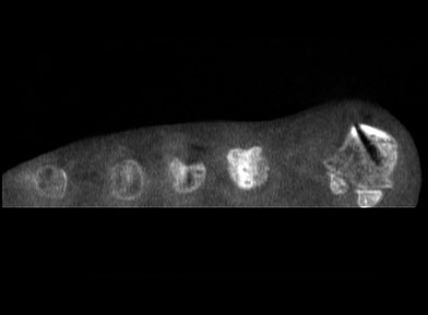
With the above background, let’s examine the contents of a weight-bearing CT image set. It permits us to visualize the target anatomy in sub-millimeter slices, from one end to the other, in all three planes. Each slice depicts just that layer, with zero interference from any tissue in front or behind it. Moreover, there is no magnification, all structures are life size. If the three original planes were not optimal for viewing a target, the planes could be tilted and re-oriented infinitely. Additionally, 3-D bone surface, skin surface and volume renderings can be generated at will, from any angle. This literally equates to thousands of clear views generated from one quick scan. You may ultimately need only a few slices to find that crucial piece of diagnostic information, but those slices are always present somewhere, in every scan, regardless of patient positioning or the beam angle.
The Value to Your Practice
Compared to the cost of a digital weight-bearing plain radiographic system (since film is on its way out anyway) weight-bearing CT costs perhaps five times as much. I would argue that the “return”, in terms of information captured, is thousands of times greater. In this particular perspective, weight-bearing CT would stand out as one of the greatest bargains we have ever been offered.
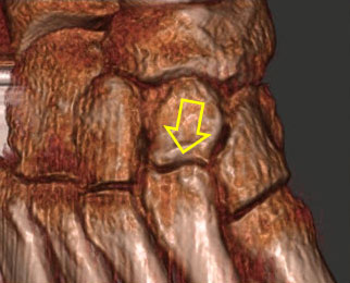
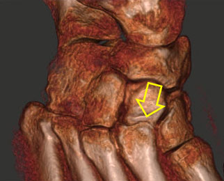
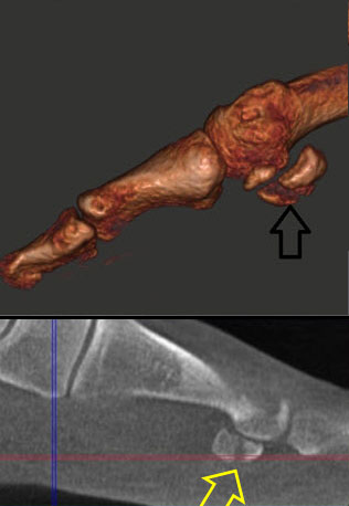
Weight-Bearing vs Non Weight-Bearing |
3D Rendering
Non Weight-Bearing |
3D Rendering
Weight Bearing |
 |
 |
|
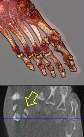
Sesamoid Fracture |
 |
 |
|
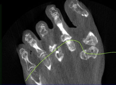
Create a custom slice through the metatarsal heads for evaluation |
 |
 |
|
As another component of the “returns”, the intangibles must be considered. Those include physician and staff time saved before, during and after treatment; better and quicker treatment outcomes, reduction in surprises and mysteries, being able to do our jobs better due to more accurate data, greater patient confidence and quicker case acceptance, etc.
Of course, the economic/monetary perspective of acquiring this device, covering both the cost and reimbursement landscapes, remains a crucial part of the analysis by necessity, and is indeed at the forefront of ongoing discussions. In the early stages, the magnitude of the “sticker price” has understandably dominated the thinking. We, as a profession, must view and analyze the full and balanced picture, which goes far beyond the price.
The Cost – How Does it Compare with Digital Radiography ?
 First, the expected life of such devices is at least 10 years, perhaps much longer. We must amortize the initial and ongoing costs over the life of the equipment, in conjunction with returns, both monetary and intangibles, to truly analyze the investment. Fortunately, we don’t need to write a check for such purchases. The equipment can be leased for up to a 10-year term, and typically it is yours for $1 at the end. For the 10-year lease example, the monthly payments are around $2,100, including taxes, in most states (less in states with no sales tax). Next, this is a fully tax deductible capital expense, so the net after tax could be around $1,400. Let’s add an allowance for service/maintenance, accreditation costs, staff time, etc., and round off the total cost at $2,000 a month. On the reimbursement side, most insurance carriers cover in-office weight-bearing CT at an average of $250 per scan. Let’s cap it at $200 after allowing for the outsourced radiologist fees, if applicable. This amounts to 10 covered scans a month.
For any group surgical practice, the above should be the proverbial “no brainer”. Let’s go a step further and contend that even for a solo surgical practice, it is hard to imagine there won’t be at least 10 patients a month who would greatly benefit from a WB CT scan.
It’s the Future – Embrace it !
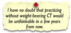 Finally, can we even imagine practicing today without X-ray? In my opinion, the jump from no X-ray to weight-bearing X-ray is dwarfed by the leap from weight-bearing X-ray to weight-bearing CT. I have no doubt that practicing without weight-bearing CT would be unthinkable in a few years from now. Are we doing justice to our patients today by depending on plain X-rays, realizing it may leave us in the dark or provide a blurred vision? In today’s age of communication, consumers are quickly learning about weight-bearing CT and its superiority. Just look at the TV news stories that ran all around the world on the weight-bearing CT scan of women’s feet in high heels, as well as such YouTube postings that went viral. Consumers are certain to demand a higher level of diagnostic imaging in the upcoming future. Can we really afford not to invest in this revolutionary technology?

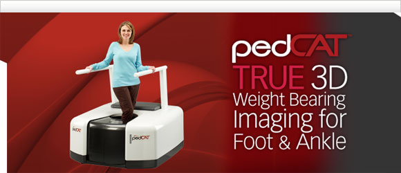
The pedCAT™, first in-office TRUE 3D weight-bearing CBCT device for the foot and ankle, puts optimal treatment planning at your fingertips for greater confidence of increased predictability in surgical outcomes. With virtually the same radiation dose as that of traditional 2D X-rays and 99% less radiation of a traditional medical CT scan, the pedCAT™ assesses clinical conditions including, but not limited to, fractures, dislocations, arthritic joints, and early detection of osteomyelitis for quicker diagnosis, evaluation and treatment planning. |
|
For more information or to request an in office demo of the pedCAT,
please contact us at 866.400.0035 or [email protected]. |