| |
Robert Frykberg,
DPM, MPH
PRESENT Editor
Diabetic Limb Salvage |
Cases From the High Risk Foot Clinic
Since our time is rather scarce and, accordingly, precious in these busy times, I thought that it would be most beneficial to simply present cases from our High Risk Foot Clinic to serve as “food for thought” in our exploration of complicated diabetic foot pathologies. As has been the format in the past, a case will be presented with a brief medical history and pertinent information including actual photographs. Since any patient can be approached in a number of different ways, it is our hope that we might stimulate blog discussion of the month’s case prior to presenting (in the next issue) the treatment that was offered. Hopefully, such a format will not only be informative, but also respect your time.
The patient this month presents a rather typical scenario for those of us who work in High Risk Foot Clinics. This patient was a 59 year-old type 2 diabetic man with diabetes duration of 15 years and controlled with insulin. He also had a history of severe coronary artery disease, congestive heart failure, mild renal insufficiency (eGFR=50), peripheral neuropathy, and peripheral arterial disease (PAD). Of particular importance, he had already succumbed to a right below knee amputation (BKA) several years prior to his presentation to us. He was referred by his primary care physician with a diagnosis of gangrene and cellulitis of the left foot. (Figure 1).
Figure 1: The patient's left foot upon presentation |
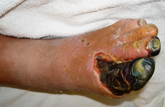 |
Our initial exam revealed absence of pedal pulses as well as neuroischemic gangrene with infection of the left foot. His Doppler signals in clinic were weakly monophasic. Prompt referral to the Vascular Laboratory for segmental limb pressures revealed an ankle-brachial index (ABI) of 0.65. Although afebrile, he was admitted for further work up and management that day. Initial lab studies indicated a leukocytosis of 12,500, hemoglobin 11.3, creatinine 1.5, serum glucose of 275 and Hemoglobin A1c of 9.3%. Gram stain showed gram positive cocci and few gram negative bacilli, but wound cultures grew out only methicillin-sensitive staphylococcus aureus. His blood cultures were negative. X-rays revealed no underlying gas or abscess, although bone was probed through the interdigital draining sinus. He was started empirically on vancomycin and piperacillin/tazobactam upon admission.
The patient’s leukocytosis rapidly resolved with antimicrobial therapy and his mild renal failure improved with hydration. But despite lack of fever, his erythema persisted. With this background information, we now need to develop a more definitive treatment plan. Points to consider are the following:
- What consultations are required?
- Is antimicrobial therapy appropriate?
- Are there other studies that we specifically need to order?
- What surgical procedures would you anticipate this patient requiring?
- Does he need another BKA to resolve his problem?
I’m sure that we’ve all seen similar patients in our clinics and perhaps we might handle them differently even in similar circumstances. Next month we will present our solutions to this person’s problem. In the meantime, we’d love to hear your thoughts on how you would approach a patient such as this.
We welcome your opinions, concerns, and suggestions. If you have an interesting case or a troubling circumstance that you would like to share with fellow PRESENT Diabetes members, please feel free to comment on eTalk.
Best regards,

Robert Frykberg, DPM, MPH
PRESENT Editor
Diabetic Limb Salvage
References:
-
Lipsky BA, Berendt AR, Cornia PB, et al. 2012 Infectious Diseases Society of America
clinical practice guideline for the diagnosis and treatment of diabetic foot infections.
Clin Infect Dis. Jun 2012;54(12):e132-173.
-
Frykberg RG, Zgonis T, Armstrong DG, et al. Diabetic foot disorders. A clinical practice guideline (2006 revision). J Foot Ankle Surg. 2006;45:S1-66.
-
Aragon-Sanchez J. Seminar review: A review of the basis of surgical treatment of diabetic foot infections. Int J Low Extrem Wounds. 2011;10:33-65.
-
Boulton AJ, Kirsner RS, Vileikyte L. Clinical practice. Neuropathic diabetic foot ulcers. N Engl J Med. 2004;351:48-55.
- Lepantalo M, Apelqvist J, Setacci C, et al. Chapter V: Diabetic foot. Eur J Vasc Endovasc Surg. 2011;42 Suppl 2:S60-74.
|