Podiatric physicians and surgeons routinely evaluate the osseous structures in their scope when performing diagnosis and treatment. After careful clinical examination where multiple diagnoses are considered, the differential list often leans to radiography for narrowing the diagnosis or confirmation of a diagnosis. Confirmation of osseous healing, whether post-injury or post-operatively (post osteotomy, arthrodesis, etc.), requires visualization of the bone. Likewise, in the study of lower extremity deformities, radiography is indispensable for comprehensive management. Aside from injury and disease states, very often baseline radiographs may be ordered on neuropathic patients, athletes, or in pediatric patients for growth evaluation. These are all reasons why most physicians who routinely treat foot and ankle conditions have in-office X-ray machines.
2D vs 3D Imaging
 Standard X-ray generators, along with the developer machines, plumbing, chemicals, and film are not inexpensive. In addition, their usage is somewhat labor intensive, and as with any machine with many moving parts, breakdowns are not infrequent. Quality of image development has inherent inconsistencies with the traditional method and this has arguably been the primary impetus for movement towards digital technology. These newer machines, along with eliminating developer chemicals from the process, have fixed armatures, special image sensors, and software that provides digital contrast enhancement. This combination has removed many of the problematic features of standard radiograph capture and development. While one can easily appreciate the difference in the capture process and quality of these images, one can also appreciate that this technique still has one severe limitation: a 3-dimensional object cannot reliably be reconstructed using a 2-dimensional radiography technique!

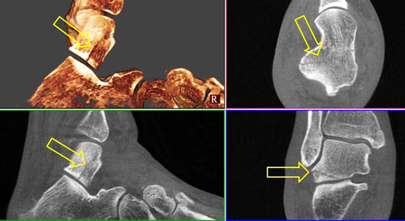
Talar Fracture
Non displaced fracture of the talus. Plain film radiography is limited in visualizing non-displaced fractures as a result of the technique. Length, orientation, and extension of fracture lines into adjacent joints is appreciated in detail with CT. |
|
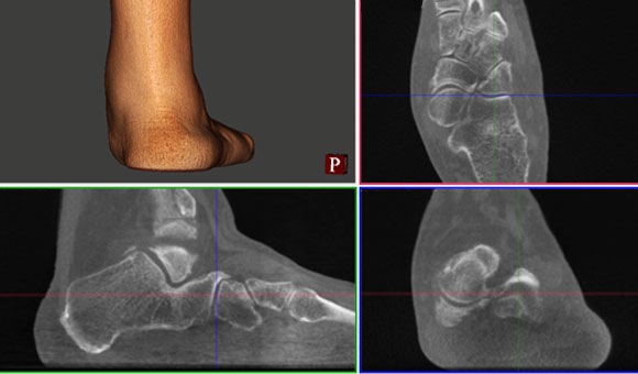
The Foot and Leg are 3D and Bear Weight
This problem, that traditionally cannot be overcome using ‘plain-film’ radiography, has led to the prescribing of more sophisticated imaging methods. Consequently, ultrasonography (US), magnetic resonance imaging (MRI), and computed tomography (CT) have been a staple in the diagnostic arsenal for decades. It is only recently that the latter method is readily available as an in-office modality for foot and ankle disorders. CT, especially when performed using a weight bearing technique, lends itself readily to hundreds if not thousands of diagnostic dilemmas, deformity scenarios, and study of poly-articular pathology. It is perplexing that a practical 3D imaging modality wasn’t adopted long ago for the lower extremity, a complicated, 3-dimensional, poly-articular structure that has a designated load-bearing kinematic purpose.
Osteochondroma
Osteochondroma as a cause of impingement. On this study the size and location of an exophytic ‘spur’ is appreciated. This is important for both understanding the pathology and for surgical planning. This study reveals that the spur is extraarticular and over the medial talar neck, so surgical approach is carried out appropriately. |
|
An Enormous Potential to Improve Podiatric Diagnosis
A staple modality in foot and ankle fracture evaluation (e.g., Lisfranc fracture/dislocations, calcaneal and pilon fractures), CT has now advanced in the evaluation of kinematics, made possible by load bearing. This dramatically extends the indications for studies where kinematics leads to pathology. For example, medial column instability can now be more fully understood, as can talo-tarsal and subtarsal subluxation, intercuneiform instability, and cuboid syndrome. Hallux limitus, one of the more compelling conditions due to its ubiquitous nature as an apex pathology (more proximal pathology often results) can now be fully understood radiographically leading to more accurate diagnosis and treatment – likewise for almost any arthrosis. Osteochondroses are not only more accurately defined for size and position, but also for their extent of contribution to joint pathology in the presence of multi-focal articular disease. Deformity after fracture healing is better appreciated specifically as it relates to weight-bearing conditions. Fractures, whether acute (e.g., sesamoid or metatarsal) or chronic (non-unions) of weight bearing bones can be understood better, as they will demonstrate measurable displacement under load, which will guide treatment more accurately. Evaluation of the infra- and posterior calcaneal spur are better understood in their anatomic structure, alignment or displacement using CT. Osteomyelitis and infectious osteitis are better managed using this modality over conventional radiographs, especially when evaluating for bone resection that may change the weight-bearing pattern (parabola in the forefoot) or midfoot. The list goes on, and every foot and ankle surgeon could probably think of hundreds of other conditions for which weight-bearing CT, if immediately available to them, would enhance their diagnostic capabilities.

Tarsal Coalition
Tarsal coalition. This patient presented with peroneal spasm, rigid flatfoot, and dorsal talonavicular beak; MRI elsewhere related no obvious coalition. This series demonstrates the coalition readily and adjacent joint degeneration. Studies like this are an essential part of preoperative planning. |
|
But at What Price?
Technology in medicine, of course, comes at a price to all who are involved: doctors, insurers, and the patients. Substantial expensive research and development was required to bring these sophisticated tools to us, but along with sophisticated tools come better diagnostic capabilities and consequently better care. The price of a wrong diagnosis, or a partial diagnosis, is far more expensive over the long run than accurate assessment the first time around. This is where the science of medicine is critical, and is why we invest in ourselves and our practices as physicians and surgeons – we know how valuable it is to make the correct diagnosis based on all the information that can be obtained. However, it makes sense to look at the complete financial picture of using in-office CT in a real-world practice environment.
Pes valgus
Pes valgus with polyarticular degeneration. Here the subtalar joint pathology is readily appreciated, as is the resultant medial column degeneration, from the hindfoot to the first metatarsophalangeal joint. Weight bearing produces the most detailed view of the joint kinematics that leads to polyarticular disease. |
|
You�ll be Using Your Weight – Bearing CT Unit a Lot
In a well-rounded foot and ankle practice, a variety of pathology is seen and treated routinely. Common conditions such as onychomycosis and tinea, uncomplicated diabetic examinations, and ingrown toenails rarely if ever warrant radiographic evaluation. However, there are other extremely common conditions where imaging is part and parcel of the diagnostic work-up. In this practice evaluation, which is fairly representative of most practices, I have compiled the top seven ICD-9 diagnosis codes (after excluding diagnoses that do not receive routine radiographs) over a six month period. In order of decreasing frequency:
729.5 - foot pain
719.47 - pain in joint, ankle and foot
726.90 - tendonitis, capsulitis, bursitis
719.97 - arthritis
728.71 - fasciitis
735.0 - bunion
735.8 - hallux limitus
Now let’s look at each code. Foot pain (729.5), is a garbage-basket type term that is as non-specific as can be possible in the lower-extremity. This code is often used when a patient presents with multiple problems, in multiple areas, or where no more definitive diagnosis can be arrived at based on the clinical exam. This is often a clinical scenario that precedes procurement of ancillary imaging and where polymodal (bone and soft tissue) CT would prove invaluable in arriving at a diagnosis. Pain in joint, ankle or foot (719.47), is commonly used when the location of pathology originates in one of the many joints of the foot or ankle. Osteochondroses (732.5 and 732.7) are often used as adjunctive codes later on or when they are large and readily apparent, although this is often not the case, at least initially. Ancillary imaging is used when flattening of the metatarsal head (first or commonly the second) is seen on plain film studies, erosive changes are seen (especially centrally in the case of the metatarsals), or a clear sign exists clinically (palpable ‘clunk’ with ROM). Arthralgia, pain with ROM, crystalline arthropathy, rheumatic flares, and even hypermobility seen with collapsing pes valgus all receive the generic 719.47 code. Of these conditions, CT can further define the diagnosis by reduction of the differential and isolation of the pathologic joint or joints. Progressive diseases such as gout and rheumatic joint diseases lend well to CT for baseline evaluation and disease advancement (719.97). Arthritis is used, when the characteristic signs of arthritis exist – chondral disappearance or flattening, periarticular spur formation, pseudocyst formation, loss of motion, joint asymmetry, etc. Diagnostic dilemmas do present in the presence of multi-focal joint degeneration and atypical presentations (e.g., isolated central tarso-metatarsal joint degeneration) that warrant further exploration. Given that a good number of arthritis cases progress to surgical intervention, CT is often used in planning and is ordered routinely prior to any procedure. Fasciitis (728.71), is generally diagnostically simple. Pain along the fascia, most often the plantar fascial bands, is extremely common in the clinical setting. Most patients respond to conservative care and thus this is not entirely an ‘interesting’ diagnosis from a radiological standpoint. However, a decent percentage do not progress with classic treatments such as NSAIDs, stretching, and orthotic therapy. In this case, one should confirm the diagnosis (to the exclusion of others such as tarsal tunnel syndrome or calcaneal/spur fracture) using ancillary imaging. Polymodal CT works extremely well for this, given that the pathology generally presents as a weight-bearing phenomenon and the study is performed in the ‘disease’ state.
Bunions and hallux limitus (735.0 and 735.8), both describe a joint disorder of the first MPJ. This is a complex joint of three articulations that is the prototype for poor imaging when standard techniques are used. Great emphasis is placed on transverse plane angulation in characterization of bunion deformity. Likewise, sagittal plane deformity is focused on in hallux limitus. It is faulty to believe that the first MPJ has such a simple description that a single radiographic parameter, regardless of the plane, can be used to dictate location, extent, and severity of the deformity in either condition. For example, weight-bearing 3-dimensional CT can demonstrate crista damage beneath the first metatarsal head in longstanding hallux valgus deformity. This parameter rarely receives attention in standard imaging techniques, but is essential to appropriate surgical planning. Concavity or convexity at the metatarsal-cuneiform articulation is also easily appreciated with this advanced image technique, an indicator of frontal plane metatarsal instability. Identification of these problems pre-operatively can lead to procedures designed to prevent recurrence of the bunion deformity after surgical correction. Frozen or retracted sesamoids, intraosseous or intra-articular pathologic foci, and proximal joint or tendon dysfunction are all imaged readily using CT, making treatment for hallux limitus through conservative or surgical means more appropriate.
Many more conditions exist that we image routinely. For example, every ulcer, with or without suspicion of osteomyelitis, is radiographed at intervals. All injuries involving blunt force or rotational components, documented fractures, and sports injuries are imaged as well. There are others, they just are not in the top seven diagnoses; but it becomes apparent how much pathology we routinely image, and how much more information can be gleaned from weight-bearing CT.
The Business Plan for Weight-Bearing CT is Compelling for Your Practice
In a practice that sees 150 patients a week, where roughly 15% are new, the potential number of new patients requiring some form of imaging is 90 a month. The top seven diagnoses comprise roughly a third of these new patients, and the argument has already been made for comprehensive imaging for any of these conditions. Somewhere between 15-20 scans a month would be ordered in this scenario. Our experience is that many existing patients are studied more extensively as well, as their complaints often present diagnostic dilemmas, so scans are not simply a modality for new patients. Because the CT scanner is in the office, a scan is readily available and takes mere minutes, so the only limitation that stands in the way of accurate anatomic/kinetic imaging is listing differentials.
The general range of ‘allowable’ payment in our area averages between $300-$400 per scan, when we looked at all payers (large commercial, Federal, and Workers Comp.) The turnkey cost of the device, including all hardware, software, installation and training is around $180,000; plus state sales taxes. The system can be leased for a term between 5 and 10 years, with $1 buyout at the end of term. The monthly payments range from around $1,980 for a 10-year lease to about $3,350 for a 5-year term. An optional service/maintenance contract costs $7,500 per year, if purchased up front for up to four additional years, which can be included in the lease. The monthly lease payments with a full 5-year service contract are around $2,300 for a 10-year term and approximately $3,900 for a 5-year term. A 7-year term is also available with intermediate figures. Accreditation costs, if Medicare billing is desired, can range from $2,600 to $7,500; depending on utilization of consulting services.
The above adds up to between 8 and 20 covered scans a month to break even, which should be a relatively low threshold for most surgical practices. We estimated that you’ll be doing around 90. At $350 per scan, this represents $25,000 additional income for your practice every month, not counting probable cash paying patients. This demonstrates the financial feasibility of in-office weight-bearing CT, not just for diagnostic purposes, but as a profit center.

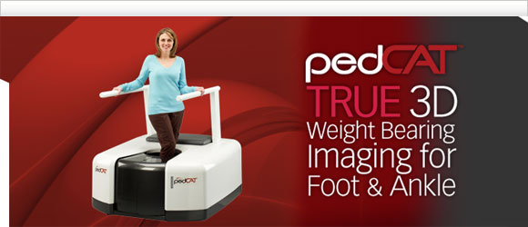
The pedCAT™, first in-office TRUE 3D weight-bearing CBCT device for the foot and ankle, puts optimal treatment planning at your fingertips for greater confidence of increased predictability in surgical outcomes. With virtually the same radiation dose as that of traditional 2D X-rays and 90% less radiation of a traditional medical CT scan, the pedCAT™ assesses clinical conditions including, but not limited to, fractures, dislocations, arthritic joints, and early detection of osteomyelitis for quicker diagnosis, evaluation and treatment planning. |
|
For more information or to request an in office demo of the pedCAT,
please contact us at 866.400.0035 or [email protected]. |