
Practice Perfect 935
A Clinical Question: What Margins are Recommended for Skin Cancers?
A Clinical Question: What Margins are Recommended for Skin Cancers?

The power of coincidence always surprises me. Recently, during resident academics, one of the residents presented the case of a patient with a skin cancer. One of our very smart attendings then asked the good question, “What size margins should you take during the surgery?” This led to an area of knowledge deficiency and further reading for the residents.

Literally the next day, I received a consultation to see a patient with malignant melanoma. Yes, the next day! One of my partners saw the patient, and we had an interesting discussion about her care. It appears the universe was telling me to write an editorial about the topic, so to avoid upsetting the universe here’s a high yield reminder about what margin sizes we should take for several common skin cancers – a podiatric service announcement, if you may!
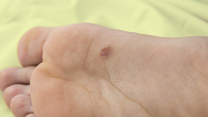
Figure 1. Malignant melanoma in situ. Courtesy Reece Everson, DPM.
Table. Skin Margins for Malignant Skin Lesions1,2,3
| Type of Malignant Skin Lesion | Risk/Characteristics | Recommended Excision Margin |
| Basal Cell Carcinoma (BCC | Low risk (small, nodular, superficial) | 4–5 mm |
| High risk (large, recurrent, aggressive subtypes, critical areas) | 5–10 mm | |
| Squamous Cell Carcinoma (SCC) | Low risk (small, well-differentiated) | 4–6 mm |
| High risk (large, poorly differentiated, recurrent, perineural invasion, critical areas) | ≥ 6 mm (up to 1 cm) | |
| Melanoma (In situ) | Localized, in situ | 0.5 cm |
| Melanoma ≤ 1 mm thick | Thin melanoma | 1 cm |
| Melanoma 1.01–2 mm thick | Intermediate thickness | 1–2 cm |
| Melanoma 2.01–4 mm thick | Thick melanoma | 2 cm |
| Melanoma > 4 mm thick | Very thick melanoma | 2 cm |


We’ll close out this podiatric service announcement with a few additional comments.
- The thicknesses stated are based on the Breslow depth classification for melanoma.4
- Recall that the margin extends beyond the actual lesion so the size of the resulting skin defect will be the size of the lesion + the margins on all sides. This may lead to a large final defect that might require plastic surgical methods of closure.
- Because the lower extremity is a unique end organ, a sizable margin of excision may require local or limb amputation in some cases.
- For melanoma, sentinel lymph node biopsy is often performed for melanomas with a thickness greater than 1 mm or for those with other high-risk features. A team approach is often helpful.
Now that we’ve discussed this topic, watch out that it doesn’t walk into your office. The universe is watching!
Best wishes.

Jarrod Shapiro, DPM
PRESENT Practice Perfect Editor
[email protected]

-
Nahhas AF, Scarbrough CA, Trotter S. A review of the global guidelines on surgical margins for nonmelanoma skin cancers. J Clin Aesthet Dermatol. 2017 Apr;10(4):37-46.
Follow this link -
Swetter SM, Tsao H, Bichakjian CK, Curiel-Lewandrowski C, Elder DE, Gershenwald JE, Guild V, Grant-Kels JM, et al. Guidelines of care for the management of primary cutaneous melanoma. J Am Acad Dermatol. 2019 Jan;80(1):208-250.
Follow this link -
Miller SJ. The National Comprehensive Cancer Network (NCCN) guidelines of care for nonmelanoma skin cancers. Dermatol Surg. 2000 Mar;26(3):289-292.
Follow this link -
Breslow A. Thickness, cross-sectional areas and depth of invasion in the prognosis of cutaneous melanoma. Ann Surg. 1970 Nov;172(5):902-908.
Follow this link

Comments
There are 0 comments for this article