
Podiatric Vein Care
Treating the Insufficient Vein: Current Modalities in Endovenous Care
Treating the Insufficient Vein: Current Modalities in Endovenous Care

In other blogs, we have identified the etiology of a bulging varicose vein, reflux and chronic vein insufficiency. If a patient presents with bulging, rope-like varicose veins, it is your responsibility to treat the underlying cause of the varicose veins first. If you were to just treat the varicose vein through sclerotherapy or an ambulatory phlebectomy, the patient would have a recurrence within a couple of years. If injection therapy is performed alone, in all likelihood, the bulging varicose vein would not go away. High concentrations of solution would be required to get a sclerosing effect on such a large bulging vein and the likelihood of trapped blood or coagulum inside the vessel leading to skin staining is exceptionally high.
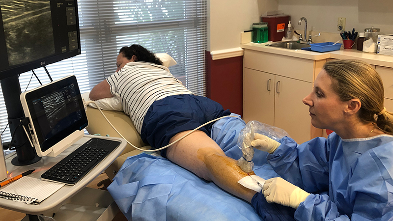
Of course, when somebody presents with bulging varicose veins, an ultrasound is used to scan the leg. Once you have identified the refluxing vein, it should be addressed through endovenous techniques. There are four common FDA approved techniques on the market; endovenous laser ablation, radio frequency (RF), VenaSeal™ which uses a medical adhesive (cyanoacrylate) and Varithena®, which is a polidocanol foam that is injected. All of these have been proven to be effective but each have their own side effects and risks.
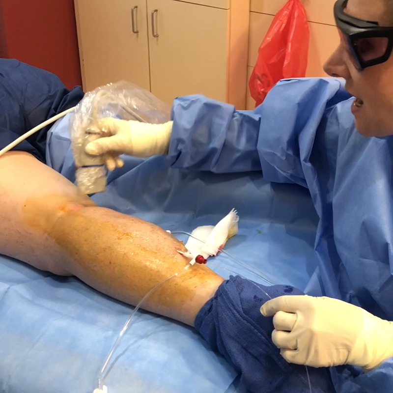
All modalities are performed under the guidance of an ultrasound. If you are not proficient in U.S. techniques, it is advisable to have an RVT or a U.S. tech assist you in the cases. If you are proficient in ultrasound maneuvering and visualization, the case can be performed alone. Endovenous laser, RF and VenaSeal are all performed by entering the vein with a needle. Through the needle, a wire is placed into the vein. The needle is removed and over the wire, a vein dilator and sheath are positioned in the vein. Once the sheath is in place, the wire and dilator are removed. If a laser is used, the fiber is placed into the sheath. Similarly, a cathode is used in RF and an adhesive guide in VenaSeal. With laser and RF, tumescent anesthetic is used. No anesthesia is required for VenaSeal. Placement is visualized through vein access, ensuring that the instrumentation is in the vessel lumen. Care is taken NOT to enter the deep system and access near a junction is positioned approximately one inch distal to the SFJ or SPJ. Varithena is performed by ultrasound-guided injections along the vein, where the foam is visualized as it travels to targeted veins.
Each modality is indicated in different types of vein refluxing portions of veins and it is imperative for the phlebologist to understand. For example, it is not recommended to perform endovenous laser in the lower portion of the leg due to proximity of the saphenous nerve and potential for nerve damage. In this instance, Varithena or VenaSeal are the procedures of choice.

Comments
There are 1 comments for this article
In response to the question that I received, "When doing evlt, is the Podiatrist allowed to treat up to the sfj?" Every state scope and laws are different. If you are uncertain, reach out to your state board to inquire. If you are interested, you may present a declaratory statement for acceptance.